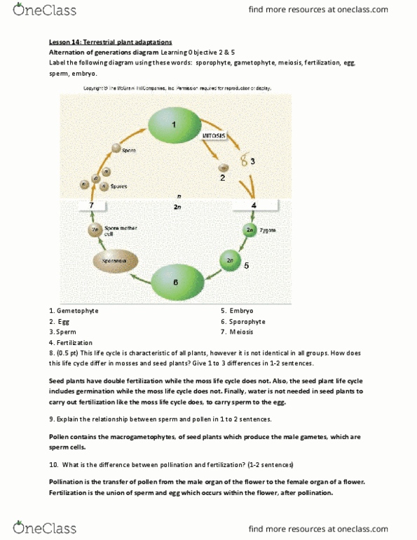
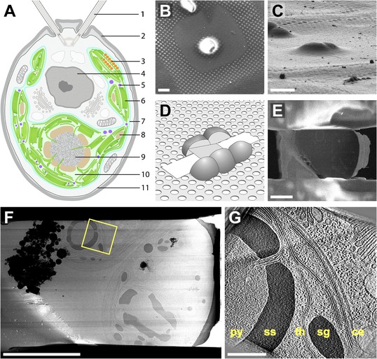
45 chlamydomonas diagram with labels
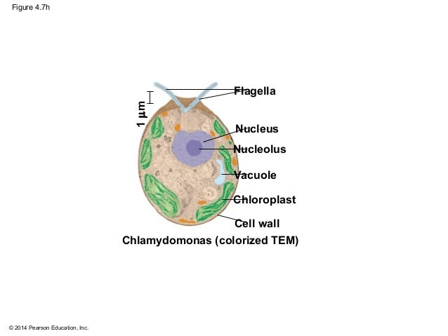
Solved: Chapter 21 Problem 24TY Solution - Chegg ISBN-13: 9780077388508 ISBN: 007738850X Authors: Sylvia S Mader Rent | Buy. This is an alternate ISBN. View the primary ISBN for: Biology 10th Edition Textbook Solutions. Biological drawings. Structure of Chlamydomonas. Learning Resources for ... Structure of Chlamydomonas: Next Drawing > Chlamydomonas is the name given to a genus of microscopic, unicellular green plants (algae) which live in fresh water. Typically their single-cell body is approximately spherical, about 0.02 mm across, with a cell wall surrounding the cytoplasm and a central nucleus.
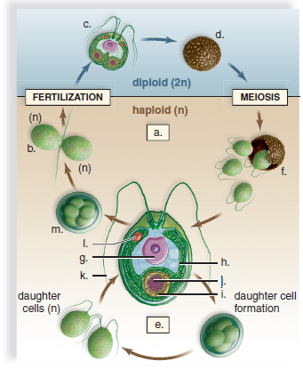
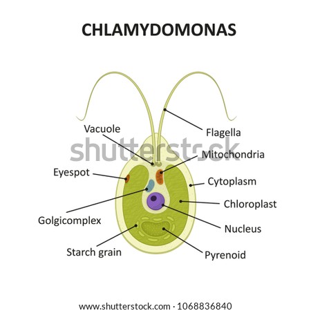
Structure of Chlamydomonas (With Diagram) | Genetics In this article we will discuss about the structure of chlamydomonas (explained with labelled diagram). The unicellular green alga Chlamydomonas is haploid with a single nucleus, a chloroplast and several mitochondria (Fig. 9.3). It can reproduce asexually as well as sexually by fusion of gametes of opposite mating types (mt + and mt - ).

Chlamydomonas diagram with labels
LABORATORY 9 - Susquehanna University Labeled diagram of Chlamydomonas. ... Chlamydomonas from culture. Cells have been stained with Lugol's Iodine, which complexes with true starch to turn black. 400X . You have slides of colonial volvocine green algae, which include Volvox, Gonium , Eudorina, ... Diagram Of Chlamydomonas With Label - Blogger Draw a labelled diagram of chlamydomonas. It is oblong or pyriform in shape. Biological drawings of protista, structure of chlamydomonas,. The anterior end has two tinsel shaped . Shipping a package with ups is easy, as you can print labels for boxes, paste them and even schedule a pickup. Diagram of Chlamydomonas angulosa... - Getty Images UNSPECIFIED - CIRCA 2003: Diagram of Chlamydomonas angulosa, Flagellated Protozoan. Drawing. (Photo by DeAgostini/Getty Images)
Chlamydomonas diagram with labels. genomebiology.biomedcentral.com › articles › 10Gene duplication and evolution in recurring polyploidization ... Feb 21, 2019 · Background The sharp increase of plant genome and transcriptome data provide valuable resources to investigate evolutionary consequences of gene duplication in a range of taxa, and unravel common principles underlying duplicate gene retention. Results We survey 141 sequenced plant genomes to elucidate consequences of gene and genome duplication, processes central to the evolution of ... › articles › nature21417Root microbiota drive direct integration of phosphate stress ... Mar 15, 2017 · a, Diagram of PSR regulation in Arabidopsis. Red and blue stripes indicate whether these mutants hyper- or hypo-accumulate Pi, respectively, in axenic, Pi-replete conditions. Describe the structure of chlamydomonas with neat labelled diagram ... answeredOct 30, 2020by Naaji(56.8kpoints) selectedOct 30, 2020by Jaini Best answer 1. Chlamydomonas is a simple, unicellular, motile fresh water algae. They are oval, spherical or pyriform in shape. 2. The cell is surrounded by a thin and firm cell wall made of cellulose. 3. The cytoplasm is seen in between the cell membrane and the chloroplast. 4. Morphology of Chlamydomonas (With Diagram) | Algae In this article we will discuss about the external morphology of chlamydomonas. Also learn about its Neuromotor Apparatus, Electron Micrograph, Palmella-Stage with suitable diagram. 1. The organism is an unicellular alga (Fig. 11). 2. The thallus is spherical to oblong in shape but some species are pyriform or ovoid. ADVERTISEMENTS: 3.
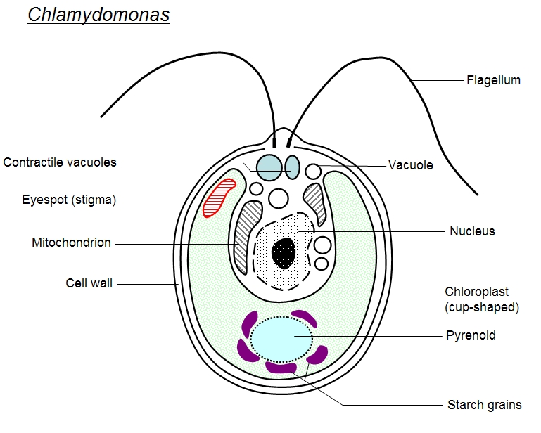
catalog.jccc.edu › coursedescriptions › biolBiology (BIOL) < Johnson County Community College - JCCC BIOL 135 Principles of Cell and Molecular Biology (4 Hours). This course is for biology majors and students planning to take additional courses in the life sciences. Subjects covered include the nature of science; the levels of organization and emergent properties of life; basic biochemistry and bioenergetics; cell structure and function; cellular reproduction; Mendelian and molecular genetics ... Solved: Label this diagram of the Chlamydomonas life cycle. | Chegg.com LearnSmart Online for Biology (10th Edition) Edit edition. This problem has been solved: Solutions for Chapter 21 Problem 24TY: Label this. diagram of the. Chlamydomonas life cycle.…. Get solutions. Get solutions Get solutions done loading. Looking for the textbook? Genetic map of the Chlamydomonas reinhardtii plastid genome ... Download scientific diagram | Genetic map of the Chlamydomonas reinhardtii plastid genome. Protein-coding regions are yellow and their exons are labeled with an "E" followed by a number denoting... Chlamydomonas reinhardtii - an overview | ScienceDirect Topics Chlamydomonas reinhardtii cells are oval shaped, c. 10 μm in length and 3 μm in width, with two flagellae at their anterior end (Figure 1). The cells contain a single chloroplast occupying 40% of the cell volume and several mitochondria. ... Diagram labeling densities in the averaged image. (B) Image average from thin sections of pf14 ...
Chlamydomonas - Meaning, Structure, Life Cycle, Function and FAQs - VEDANTU Every flagellum has two contractile vacuoles at the base. A small red eyespot can be found on the chloroplast's anterior side. Given below is the Chlamydomonas structure with labels. The Life Cycle of Chlamydomonas . Chlamydomonas Reproduction is both sexual as well as asexual reproduction. Asexual reproduction takes place by following methods: 1. Life Cycle of Chlamydomonas (With Diagram) - Biology Discussion Each daughter cell develops cell wall, flagella and transforms into zoospore (Fig. 6). The zoospores are liberated from the parent cell or zoosporangium by gelatinization or rupture of the cell wall. The zoospores are identical to the parent cell in structure but smaller in size. The zoospores simply enlarge to become mature Chlamydomonas. Chlamydomonas - Wikipedia Drawings of Chlamydomonas caudata Wille. [1] Cross section of a Chlamydomonas reinhardtii cell Light micrograph of Chlamydomonas with two flagella just visible at bottom left Chlamydomonas globosa, again with two flagella just visible at bottom left Chlamydomonas as a Model Organism - Rice University Chlamydomonas as a Model Organism. Chlamydomonas, a genus of unicellular photosynthetic flagellates, is an important model for studies of such fundamental processes as photosynthesis, motility, responses to stimuli such as light, and cell-cell recognition.C. reinhardi, the most commonly studied species of Chlamydomonas, has a relatively simple genome, which has been sequenced.

Diagram of chlamydomonas - Science - Microorganisms Friend and Foe - 9143503 | Meritnation.com
Use this labeled diagram of a chlamydomonas cell to Use this labeled diagram of a Chlamydomonas cell to address the following two questions. 32. Which of the following statements correctly identifies aspects related to photosynthesis and/or respiration? 1. Acetyl CoA is most often found in G. 2. NADPH accumulates in C. 3. ATP is found in F. 4.
35 Can You Label A Diagram Of The Alternation Of Generations Life Cycle_ - Labels Database 2020
Draw a neat labelled diagram. Chlamydomonas - Shaalaa.com Draw a neat labelled diagram. Chlamydomonas . Maharashtra State Board HSC Science (General) 11th. Textbook Solutions 8018. Important Solutions 19. Question Bank Solutions 5546. Concept Notes & Videos 432. Syllabus. Advertisement Remove all ads. Draw a neat labelled diagram. ...

VIRIDIPLANTAE
Asymmetric properties of the Chlamydomonas reinhardtii cytoskeleton ... The C. reinhardtii eyespot. (a) A diagram illustrating asymmetric localization of the eyespot relative to the cytoskeleton. Two flagella and four microtubule rootlets extend from a pair of basal bodies at the anterior end of the cell; both the mother basal body (small black oval) and the daughter basal body (small gray oval) are associated with a four-membered rootlet (M4 or D4) and a two ...

AP Biology-Ch.6 A Tour of the Cell
Structure of Chlamydomonas (With Diagram) | Chlorophyta In this article we will discuss about the structure of chlamydomonas with the help of suitable diagrams. Chlamydomonas is unicellular, motile green algae. The thallus is represented by a single cell. It is about 20 p,-30|i in length and 20 µ in diameter. The shape of thallus can be oval, spherical, oblong, ellipsoidal or pyriform.
Chlamydomonas – Parts Labeled | Radesaal the science group
Eye Diagram With Labels and detailed description - BYJUS A brief description of the eye along with a well-labelled diagram is given below for reference. Well-Labelled Diagram of Eye The anterior chamber of the eye is the space between the cornea and the iris and is filled with a lubricating fluid, aqueous humour. The vascular layer of the eye, known as the choroid contains the connective tissue.
Native architecture of the Chlamydomonas chloroplast revealed by in situ cryo-electron ...
› pmc › articlesActin and Actin-Binding Proteins - PMC A seed was first decorated with myosin heads and then allowed to grow bare extensions. Elongation was faster at the barbed end than at the pointed end. (B) Diagram showing the rate constants for actin association and dissociation at the two ends of an actin filament. The pointed end is at the top and the barbed end is at the bottom.
Schematic diagram of chlamydomonas | Algae - unclassified | Science textbook, Diagram, Green algae
Chlamydomonas: Position, Occurrence and Structure (With Diagrams) Chlamydomonas is unicellular, motile green algae. The thallus is represented by a single cell. It is about 20 p,-30|i in length and 20 µ in diameter. The shape of thallus can be oval, spherical, oblong, ellipsoidal or pyriform. The pyriform or pear shaped thalli are common, they have narrow anterior end and a broad posterior end (Fig. 1).

Metabolic network reconstruction of Chlamydomonas offers insight into light‐driven algal ...
› science › articleSingle-cell mass spectrometry - ScienceDirect May 11, 2022 · The labels are designed to ensure that (i) the total mass of each TMT label (reporter and linker groups) is identical, and (ii) the reporter groups have 18 different masses. Thus, a given peptide ion that is tagged with different labels will have identical masses for m / z selection and ion fragmentation, resulting in abundant sequence ions for ...

Lab Practical 101 at University of Puerto Rico - Cayey - StudyBlue
Biology Diagram Of Chlamydomonas - Which One Of The Following Is A ... Draw A Labeled Diagram Of Chlamydomonas Snapsolve from wb-qb-sg-oss.bytededu.com I tell you about how can we draw labelled diagram of chlamydomonas in . Science, 8th standard text book, ktbs. The thallus is represented by a single cell. Draw chlamydomonas step by step drawing easy to draw#sudhakararts#biological sciences#drawing.
Solved: Label this diagram of the Chlamydomonas life cycle. | Chegg.com
› de › financeFinances in Germany - Expat Guide to Germany | Expatica Learn everything an expat should know about managing finances in Germany, including bank accounts, paying taxes, getting insurance and investing.
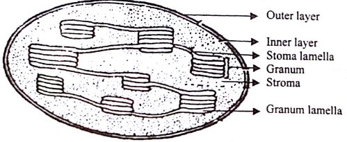
Describe the Structure of Chloroplast - QS Study
Spirogyra Labelled Diagram Spirogyra (common names include water silk, mermaid's tresses, and blanket weed) is a genus of filamentous charophyte green algae of the order Zygnematales, named for the helical or spiral arrangement of the chloroplasts that is characteristic of the genus. Draw a labelled diagram of Spirogyra. 51 Differentiate between flying lizard and bird.
Euglena Cartoons, Illustrations & Vector Stock Images - 110 Pictures to download from ...
Animal Cells: Labelled Diagram, Definitions, and Structure Only present in lower plant forms (e.g. chlamydomonas) Present in all animal cells: Chloroplast: Plant cells have chloroplasts to synthesize their own food. Absent: Plasma Membrane: Cell wall and a cell membrane: Only cell membrane: Flagella: Present in some cells (e.g. sperm of bryophytes and pteridophytes, cycads and Ginkgo)
Biology in Focus Chapter 4
How to make label Diagram of chlamydomonas - YouTube watch: "How to make thumbnail our you tube videos Hindi /urdu haris by #Top2utv" ...

Dynein and its arrangement in sea urchin sperm flagella. (a) Sequence... | Download Scientific ...
Chlamydomonas | Facts, Structure, Life Cycle, & Classification Chlamydomonas, genus of biflagellated single-celled green algae (family Chlamydomonadaceae) found in soil, ponds, and ditches. Chlamydomonas species can become so abundant as to colour fresh water green, and one species, C. nivalis, contains a red pigment known as hematochrome, which sometimes imparts a red colour to melting snow. The cells of most Chlamydomonas species are more or less oval ...

Labelled Diagram Of Chlamydomonas - Top Label Maker
› microorganisms-friend-and-foeMicroorganisms: Friend and Foe Class 8 Extra Questions ... Oct 11, 2019 · Pull out a gram or bean plant from the field. Observe its roots. You will find round struc¬tures called root nodules on the roots. Draw a diagram of the root and show the root nod¬ules. Answer: Question 2. Collect the labels from the bottles of jams and jellie on the labels. Answer: Do it yourself. Question 3. Visit a dcotor.
Structure Chlamydomonas Stock Vector (Royalty Free) 1068836840 - Shutterstock
Chloroplast Structure and Function in detail with Labelled Diagram The chloroplasts are the cell organelles which consist of these pigments. The 3 types of pigments present in plants are chlorophyll, carotenoids, and anthocyanins. Chlorophyll imparts the green color to plants. Plastids are membrane-bound cytoplasmic organelles that can be found in the cells of plants and algae.
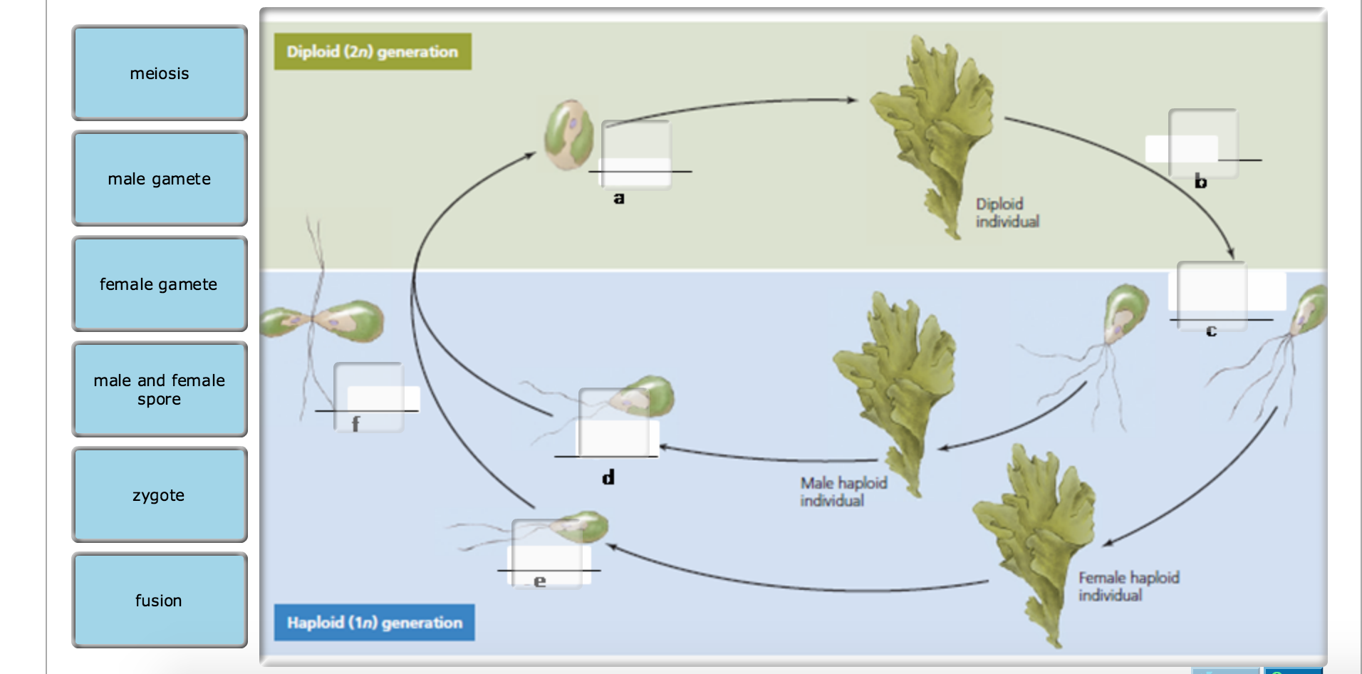
32 Can You Label A Diagram Of The Alternation Of Generations Life Cycle_ - Labels Database 2020
The Chlamydomonas Flagellum as a Model for Human ... - ScienceDirect (The images and diagrams of Chlamydomonas central pairs are from Lechtreck and Witman, 2007; ... Although the antibody labeled internal membranes, the strongest labeling, and the only labeling on the surface of the cell, was in the cilium. These results demonstrated that polycystin-2 is displayed specifically on the primary cilium.
Post a Comment for "45 chlamydomonas diagram with labels"